New · since 1 October 2025
Equine CT scanner at Les Bréviaires
New equipment at the service of your horse.
Since 1 October 2025, the Clinique équine des Bréviaires has been equipped with a new latest-generation CT scanner, allowing more accurate diagnoses and better care for your horse. Whether your horse is already followed by our team or by an outside veterinarian, this examination is available to all horse owners who wish to benefit from this advanced imaging technology.
The technology
How does the scanner work?
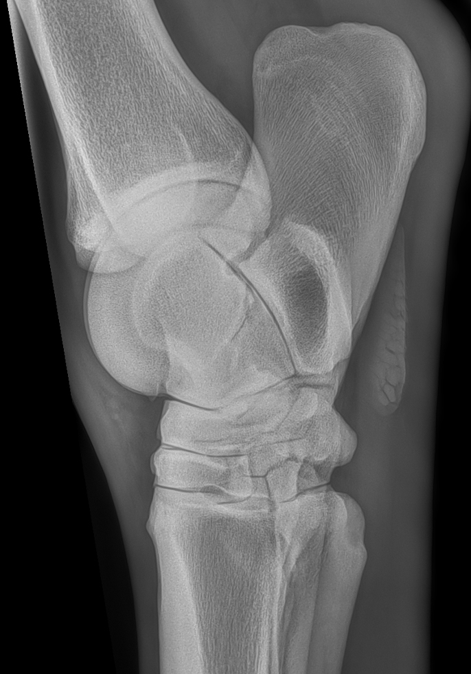
A CT (computed tomography) scan is an imaging technique that uses X-rays, like radiography. Unlike radiography, which produces two-dimensional images, CT is a three-dimensional imaging technique. It relies on the continuous rotation of an X-ray generator and a detector system, which acquires several dozen images per second.
This technology makes it possible to analyse the images in every plane by slicing the horse's anatomy into very thin sections, and also to produce three-dimensional reconstructions.
Our equipment
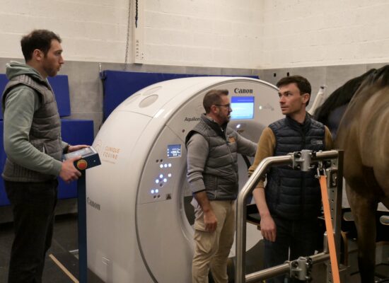
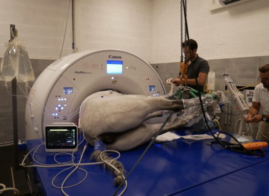
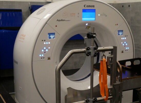
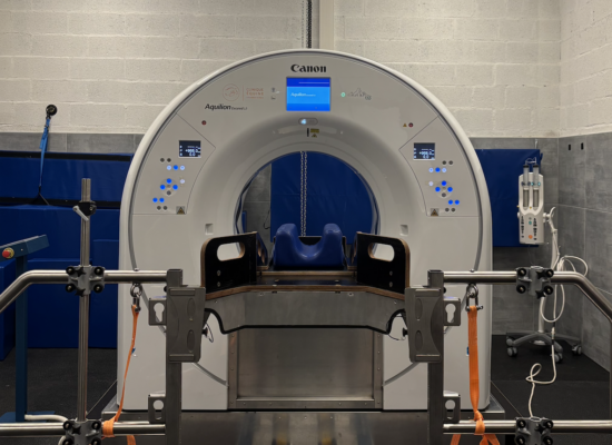
Canon Aquilion Exceed LB scanner
01
Eleva CT platform
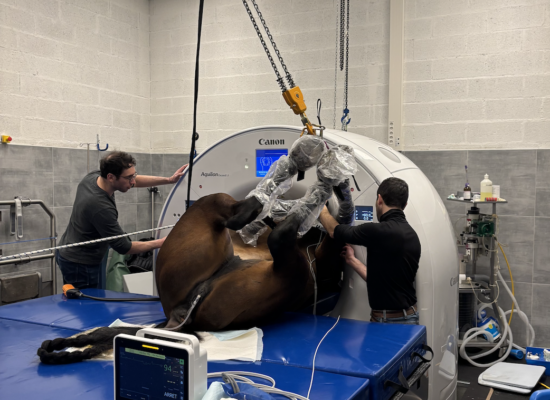
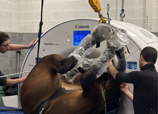
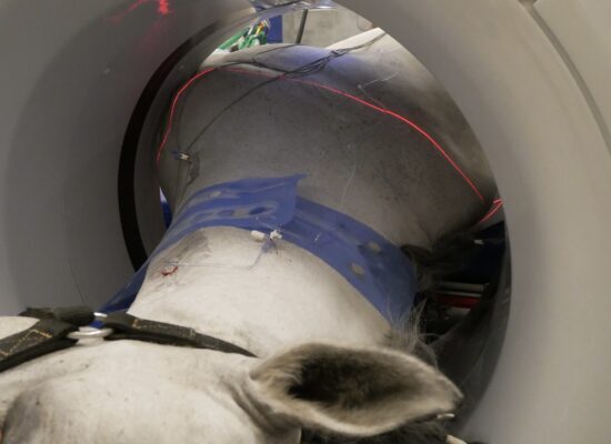
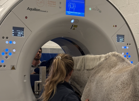
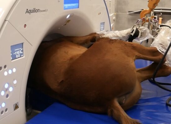
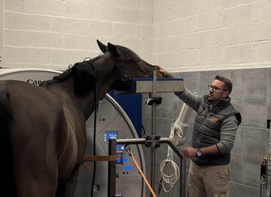
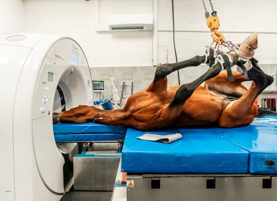
Unlike conventional scanners where the horse stays still, our platform rises, lowers and moves, for standing examinations under light sedation or lying examinations under general anaesthesia.
02
Contrast CT
Contrast is injected directly from the reading room, simultaneously with image acquisition: a service offered by only 2 clinics in France.
03
Dual Energy
A technology on which our teams are carrying out research, with the aim of better showing bone oedema on CT.
90 cm
gantry diameter
10 to 20 s
to scan a large region
Dip. ECVDI
scans read by a specialist
Thanks to its wide 90 cm gantry and its many detectors, this scanner can image large regions of the horse (neck, shoulders, stifles, pelvis) in only 10 to 20 seconds. All examinations performed at the clinic are carried out and interpreted by Dr Alexandre Charles, a veterinary imaging specialist certified by the European College of Veterinary Diagnostic Imaging (Dip. ECVDI). A detailed report is always sent to you in the days following the examination.
Your imaging specialist
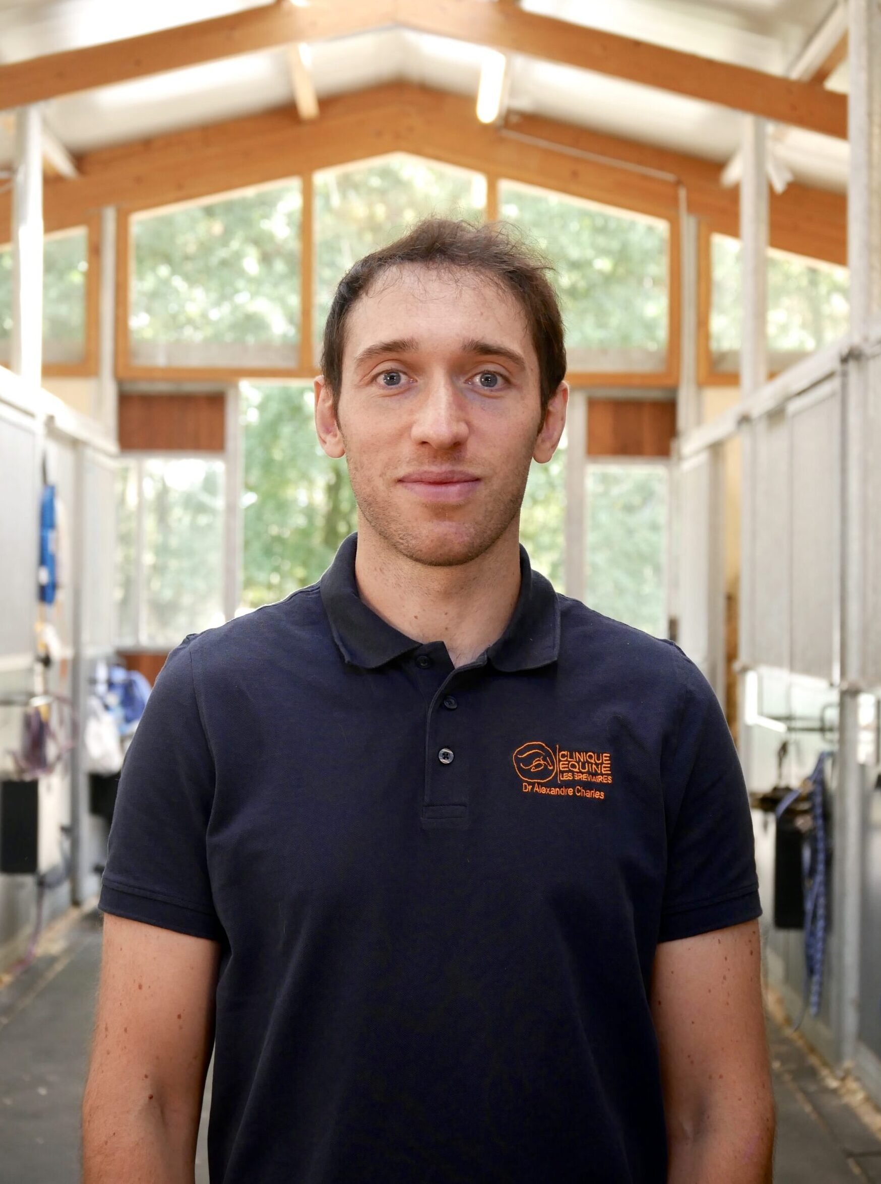
Dr Alexandre Charles
Veterinary imaging specialist · Dip. ECVDI
Alexandre graduated from ENVA in 2018 and completed a one-year internship in equine clinical practice at the school, followed by an assistantship at CIRALE. After a period in practice at the Clinique équine des Bréviaires, he joined the imaging team of the University of Liège veterinary teaching hospital, where he continued his training with a specialised internship and then a residency in imaging. Certified by the European College of Veterinary Diagnostic Imaging (ECVDI) in 2025, he remains a part-time senior clinician at the university while developing his activity with several clinics in France.
He joined the Bréviaires clinic in September 2025, at the launch of the scanner project, and trained the team to use it. There he performs the cross-sectional imaging examinations and interprets them, on site and remotely, and develops several research projects in this field.
When to use it
Indications
01
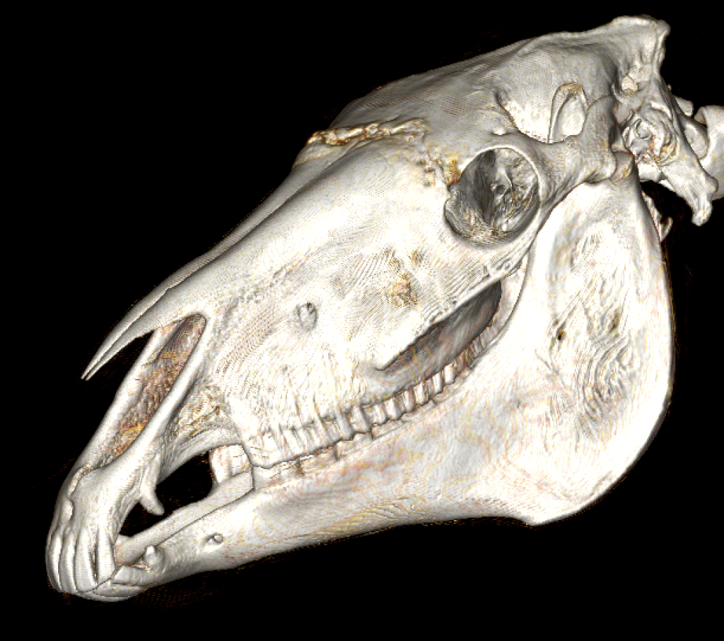
Head examination
For investigating dental and sinus disease, head-shaking, disorders of the upper respiratory tract or of the central nervous system.
02
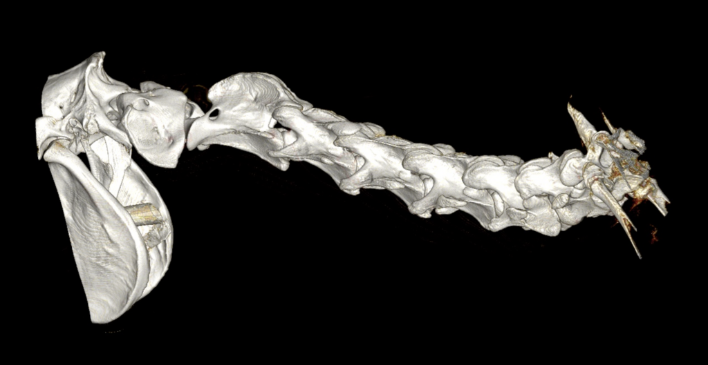
Cervical spine examination
Which can be supplemented by a myelogram, for investigating ataxia, neck pain or forelimb lameness of axial origin.
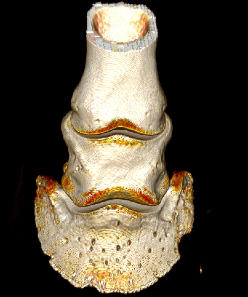
03
Forelimb and hindlimb examination
For lameness, fractures or the investigation of a mass.
04
Chest and abdomen examination
For investigating respiratory disorders and gastrointestinal and urogenital disease.
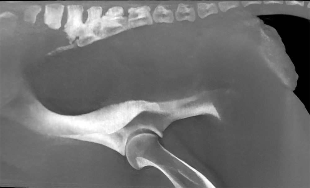
05
Pelvis examination
Depending on the size of the animal, for investigating high lameness and lack of propulsion and/or engagement of the hindquarters.
These examinations may include procedures such as contrast injection to help visualise musculotendinous and joint structures. The indications and associated procedures are discussed beforehand with the referring veterinarian and/or the owner, depending on the clinical problem.
Book
How to book an appointment
Your horse is followed by the clinic
If your horse is followed by a veterinarian at the Bréviaires clinic, get in touch with them so they can examine your horse and guide you towards the most appropriate type of examination.
You are referred by an outside veterinarian
You can make an appointment with our reception team. An examination request form to complete will be sent to your veterinarian, who may be contacted by phone if necessary to understand your horse's problem as fully as possible.
Examination day
How the examination goes and your results

Standing CT scan
Without general anaesthesia
Depending on the type of examination, your horse can be dropped off at the clinic on the same day and go home once the examination is over.
Lying CT scan
Under general anaesthesia
Examinations under general anaesthesia often require the horse to arrive the day before, when a pre-anaesthetic examination is carried out. You are contacted once the horse has woken up, and discharge from hospital is scheduled for the following day.
Results
A detailed report
Image acquisition and interpretation are supervised by a specialist veterinarian certified by the European College of Veterinary Diagnostic Imaging (Dip. ECVDI). The referring veterinarian is contacted the same day to discuss the results and your horse's management. An imaging report is sent to the owner and to the veterinarian in the days that follow.

Prices
Our prices
Feel free to contact us by phone or email to get an accurate quote.
Head scan (with or without contrast)
Between €1,100 and €1,400 incl. VAT
Standing limb scan
From €550 incl. VAT
Limb scan under general anaesthesia
From €1,400 incl. VAT
Cervical spine scan
Between €1,500 and €2,000 incl. VAT